What is “scanning”, and what do we mean when we talk about scanning fossils? Modern scanning technology has transformed palaeontology in recent years. The ability to non-destructively image internal features, capture detailed shape of very large, very small or remote specimens, digitise fossils for complex analyses, as well as share digital data easily, means entire new avenues of research and possibilities for collaboration are opening up.

The palaeontology team at Flinders University routinely use various scanning techniques (and associated data processing and visualisation software) for their research, outreach and education programs using methods detailed below. If you have a specimen that you think you might want scanned and would like to learn more about how this works, then please feel free to contact the VAMP team.
Computed Tomography (CT)

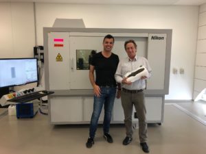
A Computed Tomography (CT) machine, sometimes also known as CAT, takes a series of X-ray images and combines them to create cross-sectional images (slices) of the object. These slices are then combined into a “stack” to create a 3-D dataset. As a CT works on the principles of radio-density, internal structure as well as external features are captured. MicroCT is simply CT but with higher resolution of the images captured.

For more information about using the Tonsley XCT Facility:  https://www.flinders.edu.au/microscopy/materials-characterisation/large-volume-micro-ct-system

Synchrotron Tomography

A synchrotron accelerates electrons close to the speed of light, then releases this extremely bright light into various beamlines for research or medical use. Synchrotron light is incredibly bright (a million times brighter than the sun!) and can be generated across the electromagnetic spectrum from infrared to X-rays. Synchrotron imaging uses the same principles as CT imaging, that is imaging using X-rays and converting multiple radiographs of an object into 3-D datasets.

Australian Synchrotron ANSTO: https://www.ansto.gov.au/facilities/australian-synchrotron

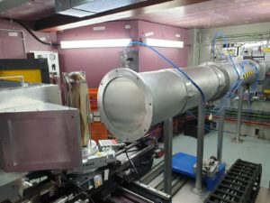
Neutron Tomography

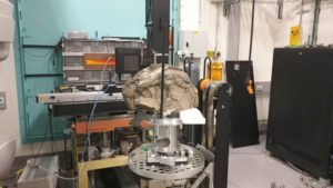
Unlike X-rays, which rely on electrons and attenuate depending on the density of an object, neutron imaging, as its name suggests, relies on neutrons. This means that how an object is imaged is not related to its density (as in X-rays) but instead on the chemical composition of the materials within the sample (and other neutron attenuation properties).

Neutron Beam – DINGO at ANSTO https://www.ansto.gov.au/facilities/australian-centre-for-neutron-scattering

Photogrammetry & LIDAR

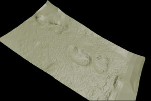
Photogrammetry is an extremely versatile tool whereby photos can be used to construct a three-dimensional image. Software identifies points shared across multiple photos and uses this to triangulate their positions. This then leads to the generation of a point cloud, sometimes consisting of 100s of thousands or even millions of individual points. From this point cloud a 3D mesh is then generated and the photo texture is mapped onto the mesh, resulting in a highly accurate 3D render of the original object. This low-cost method is useful in lab-based settings but can be critical for field-based studies where data capture using more sophisticated scanning techniques isn’t possible: the only gear you need to carry with you is a camera and tripod.

LIDAR scanning works in a similar way to photogrammetry in that a point cloud is generated but instead of photographs, lasers shoot out and are reflected back from the scanned surfaces. This produces distance measurements to surrounding objects which can then be used to assemble a point cloud. Depending on the scanner used, this can lead to a much higher resolution model than can be generated from photogrammetric methods and it is often employed in field situations for generation of site- or even landscape-level renders.

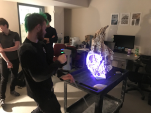
Surface Scanning

Structured light scanners work by projecting a pattern onto the object usually with a centre projector. The pattern becomes distorted when projected on to the object, which is then picked up with the side cameras which use these distortions to calculate the distance and shape of the object. This then generates a point cloud replicating the surface that was scanned. These point clouds can then be meshed and exported as a 3-D object.